
Physical Therapy Approaches to Lower Extremity Problems: A Comprehensive Review of the Hip, Knee, and Ankle
1. Introduction
The lower extremity is the cornerstone of the human movement system. Many functions, such as daily living activities, athletic performance, and postural control, depend on the coordinated work of the hip, knee, and ankle joints. Pathologies in these areas can lead not only to local pain but also to functional impairments that affect the entire kinetic chain.
Physical therapy offers a multidisciplinary approach to managing such problems. Musculoskeletal assessments, manual therapy techniques, exercise prescriptions, and patient education form the cornerstones of treatment. This article will examine physical therapy approaches to lower extremity problems in light of the scientific literature, addressing anatomical, pathological, and therapeutic aspects specific to the hip, knee, and ankle.
2. Hip Problems and Physical Therapy
2.1 Anatomical and Biomechanical Structure
The hip joint is a ball-and-socket type synovial joint located between the femoral head and the acetabulum. It is stabilized by strong ligaments and muscles. Muscles such as the gluteus maximus, medius, and minimus play a critical role in both movement and postural control.
2.2 Common Pathologies
• Trochanteric Bursitis: May develop as a result of overuse or weakness of the gluteal muscles. It is characterized by lateral hip pain.
• Gluteal Tendinopathy: Particularly in the gluteus medius and minimus tendons.
• Femoroacetabular Impingement (FAI): Pain and limited movement occur as a result of abnormal contact between the femoral head and acetabulum.
2.3 Physical Therapy Approaches
• Correcting Muscle Imbalances: Activation of the gluteal muscles is recommended, especially in conjunction with the VMO.
• Mobilization Techniques: Anterior hip glide mobilization is effective for internal rotation limitations.
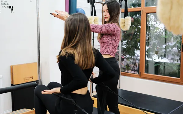
• Functional Exercises: Exercises such as step-downs and single-leg squats increase both strength and balance.
3. Knee Problems and Physical Therapy
3.1 Anatomical Structure
The knee joint is located between the femur, tibia, and patella. Stability is provided by ligaments (ACL, PCL, MCL, LCL) and surrounding muscles. Proper alignment of the patella is related to the balance of the quadriceps and hamstring muscles.
3.2 Common Pathologies
• Anterior Cruciate Ligament (ACL) Injuries: Usually occur as a result of sudden changes in direction or trauma.
• Patellofemoral Pain Syndrome: Develops as a result of misalignment between the patella and femur.
• Meniscus Lesions: Meniscus tears are common during rotational movements.
3.3 Rehabilitation Protocols
• Early Mobilization: Passive and active range of motion exercises.
• Proprioceptive Exercises: BOSU, balance board, standing on one leg.
• Functional Strength Training: Lunge, step-up, squat variations.
4. Ankle Problems and Physical Therapy
4.1 Anatomical Structure
The ankle is located between the talus, tibia, and fibula. Stability is provided by the lateral and medial ligaments.
4.2 Common Pathologies
• Lateral Sprains: Result from outward force on the ankle joint.
• Tendinopathies: Commonly seen in the peroneal and posterior tibialis tendons.
• Plantar Fasciitis: Characterized by inflammation of the plantar fascia in the sole of the foot.
4.3 Treatment Approaches
• Mobilization Techniques: Posterior talus glide, increased dorsiflexion.
• Balance and Proprioception: Standing on one foot, dynamic balance exercises.
• Stretching and Loading Strategies: Stretching the gastrosoleus complex, plantar fascia stretching.
5. Kinetic Chain and Holistic Approach
The joints of the lower extremity are interconnected. Limited ankle mobility can increase valgus moments at the knee; hip compensation can lead to gluteal inhibition.
5.1 Compensation Mechanisms
• Ankle dorsiflexion deficiency → excessive knee flexion
• Hip abductor weakness → knee valgus
• Pelvic instability → gait abnormalities
5.2 Functional Assessment
• Squat Analysis: Hip, knee, and ankle alignment are observed.
• Gait Analysis: Step length, weight-bearing pattern, balance.
6. Clinical Case Examples and Exercise Protocols
6.1 Case 1: Rehabilitation Following ACL Reconstruction
1st week: passive mobilization, quadriceps isometric
Week 4: Balance board, mini squats
Week 8: Lunges, step-ups, beginning to run
6.2 Case 2: Patient with Plantar Fasciitis
• Stretching exercises: plantar fascia, gastrosoleus
• Foot mobilization
• Foot mobilization
• Balance exercises: barefoot BOSU
6.3 Case 3: Gluteal Tendinopathy
• Hip abductor activation
• Step-down exercise
• Pelvic stability exercises
7. Discussion and Conclusion
Physical therapy for lower extremity problems requires an approach that addresses not only local symptoms but the entire movement chain. The interaction between the hip, knee, and ankle plays a critical role in treatment planning. The literature emphasizes the effectiveness of functional exercises and proprioceptive exercises.
This article aims to provide physical therapists and healthcare professionals with a holistic perspective on lower extremity rehabilitation. Future research should examine the long-term effects of individualized exercise prescriptions.
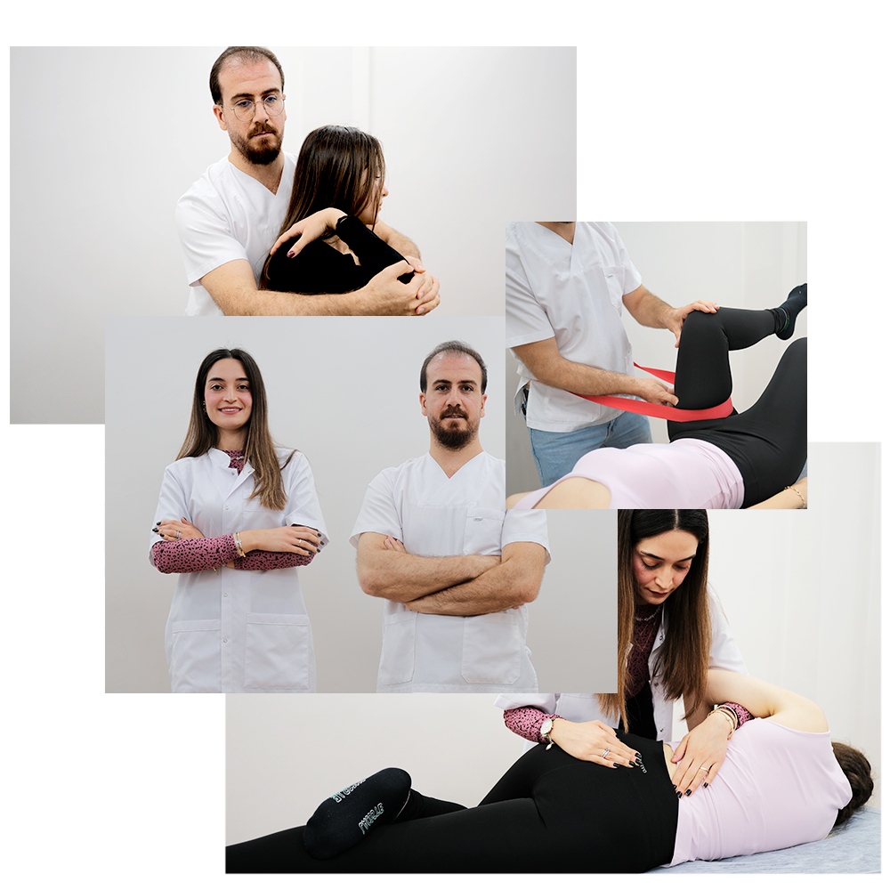
Primer Fizyoterapi Merkezini diğerlerinden ayıran şey bugüne kadar edindiğimiz saygın ve lider sağlık kuruluşu olmamız ve kişiye özel hasta-fizyoterapist deneyimi sunmamızdır.
Hemen aşağıdaki butona tıklayıp Primer Fizyoterapi Merkezini arayarak randevunuzu alabilirsiniz.
Randevu Al
